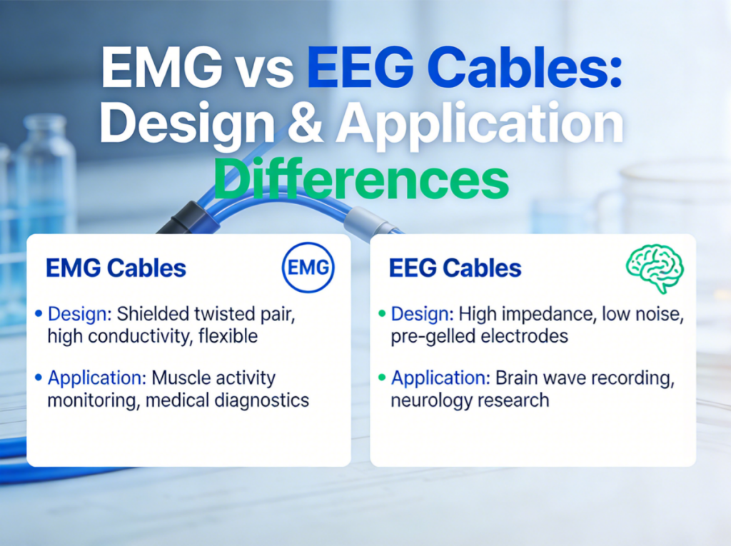
EMG (Electromyography) and EEG (Electroencephalography) cables are both critical for bioelectrical signal monitoring in clinical, surgical, and research settings—but their engineering design, performance requirements, and real-world applications differ drastically. While both transmit vital biological signals, EMG cables track muscle activity, and EEG cables capture brain activity, each demanding specialized structural and functional features to ensure accurate, reliable data collection.
For clinicians, medical device engineers, and lab researchers, understanding these core differences is essential to selecting the right cable assembly for your monitoring system. This guide breaks down the key distinctions between EMG and EEG cables—from signal characteristics and electrode design to structural engineering and ideal use cases—so you can make informed decisions for your neuromuscular or neurological monitoring needs.
What Are EMG Cables?
An EMG cable is a medical-grade cable assembly engineered exclusively to transmit electrical signals generated by muscle contraction and activity. Unlike EEG cables, which focus on the brain’s faint neural activity, EMG lead wires are designed to detect the stronger electrical responses of skeletal and smooth muscles, making them a staple in neuromuscular diagnostics and surgical monitoring.
Core Characteristics of EMG Cables
- Shorter lead lengths for direct muscle contact
- Tolerance for higher signal amplitudes (muscle signals are far stronger than brain signals)
- Compatibility with needle or surface EMG electrodes
- Configurable for bipolar or monopolar monitoring setups
- Optimized for flexibility to accommodate patient movement during testing or surgery
Primary Applications of EMG Cables
EMG cables are tailored for settings that require monitoring of muscle and motor nerve function, with key use cases including:
Neuromuscular Diagnostics
Clinical evaluations of muscle response, nerve conduction, and neuromuscular disorders (e.g., muscular dystrophy, carpal tunnel syndrome) in neurology and orthopedics clinics.
Intraoperative Neuro Monitoring (IONM)
Critical real-time monitoring of motor nerve function during surgical procedures (e.g., spinal, orthopedic, or neurological surgery) to prevent nerve damage.
Rehabilitation & Physical Therapy
Surface EMG monitoring to track muscle activation, measure recovery progress, and guide targeted muscle training for patients with injuries or mobility impairments.
Research Laboratories
Biomechanics and sports science studies focused on muscle activation patterns, movement analysis, and neuromuscular physiology.
In all these applications, EMG cables balance signal reliability with the flexibility needed to work with moving patients and dynamic testing environments.
EMG vs EEG Cables: Critical Comparative Overview
The fundamental difference between EMG and EEG cables stems from the signals they transmit: EMG cables handle stronger muscle signals, while EEG cables capture the brain’s ultra-faint microvolt-level neural activity. This translates to distinct design, performance, and electrode requirements, summarized in the table below:
| Feature | EMG Cable | EEG Cable |
| Signal Type | Muscle electrical activity | Brain cortical electrical activity |
| Signal Amplitude | High (millivolt level) | Very low (microvolt level) |
| Noise Sensitivity | Moderate | Very high (easily disrupted by EMI) |
| Electrode Type | Needle / surface electrodes | Gold-plated cup / Ag/AgCl electrodes |
| Configuration | Bipolar / monopolar | Multi-channel (for full-brain mapping) |
| Core Priority | Flexibility, electrode stability, motion resistance | Shielding, low impedance, noise rejection |
Key Takeaway: EEG cables demand extreme precision in shielding and impedance control to protect faint brain signals, while EMG cables prioritize flexibility and electrode contact stability for monitoring moving muscles.
Structural Design: Key Engineering Differences
Beyond core signal characteristics, EMG and EEG cables feature specialized structural designs to meet their unique performance needs—from impedance control and shielding to electrode termination and cable construction.
Impedance Requirements
- EEG Cables: Require strict, stable low impedance (typically <5kΩ) to prevent distortion of the brain’s ultra-faint microvolt signals. Even minor impedance fluctuations can render EEG data unusable, so tight manufacturing control is non-negotiable.
- EMG Cables: Tolerate slightly higher and more variable impedance, as muscle signals are stronger and less prone to distortion. The primary impedance focus is on stable electrode contact (rather than cable itself) to ensure consistent signal transmission.
Shielding & Noise Control
Signal interference (EMI) is a challenge for both, but the approach to shielding differs based on noise sensitivity:
- EEG Cables: Utilize multi-layered noise reduction, including twisted pair conductor structures, full cable shielding, and even multi-channel individual shielding—critical for blocking EMI in clinical or lab environments with medical equipment, lights, or electronics.
- EMG Cables: Prioritize compact, flexible shielding (rather than heavy multi-layered design) to support patient movement. Shielding is still important for surgical settings (high EMI from OR equipment) but is balanced with cable flexibility.
Electrode Configuration & Termination
Electrode type directly shapes cable termination design, as the cable must form a secure, low-resistance connection with the electrode:
- EMG Cables: Terminated for bipolar/monopolar needle or surface electrodes—small, compact connectors that fit the direct-contact nature of muscle monitoring. Needle electrode compatibility requires durable, fine-gauge lead wires.
- EEG Cables: Terminated for gold-plated cup or Ag/AgCl electrodes (the clinical standard for brain monitoring), with multi-channel connectors to support the 16, 32, or 64-channel setups used for full-brain EEG mapping.
Common Engineering Challenges for EMG & EEG Cables
While their design challenges vary, both EMG and EEG cable assemblies face shared and unique engineering hurdles—especially in high-demand clinical and surgical environments. The most common issues include:
- Signal interference: EMI from medical equipment (ORs, ICUs) disrupts faint EEG signals and can degrade EMG data in complex settings.
- Connector mismatch: Incompatible cable-to-monitor connectors cause setup delays and signal loss (a universal pain point for both cable types).
- Cable breakage: Weak overmolding at cable-connector junctions fails under repeated use or patient movement (EMG cables are particularly prone due to flexibility demands).
- Multi-channel instability: EEG’s multi-channel setups face signal crosstalk, while EMG’s bipolar/monopolar configs require consistent electrode contact.
The solution to all these challenges: partnering with an experienced, certified medical cable manufacturer with specialized expertise in both EMG and EEG cable design—one that prioritizes structural optimization, strict quality control, and system compatibility.
How to Choose: EMG or EEG Cable?
Selecting the right cable assembly boils down to your monitoring target and application environment—follow these key considerations to make the optimal choice:
- Define your monitoring target: Choose EMG cables for muscle/motor nerve activity; EEG cables for brain/neural cortical activity.
- Assess signal sensitivity needs: Prioritize EEG cables for ultra-low microvolt signals (requiring maximum shielding/impedance control); EMG cables for higher millivolt muscle signals (prioritizing flexibility).
- Match to the application environment: EMG cables are ideal for dynamic settings (surgery, physical therapy, patient movement); EEG cables excel in static/controlled settings (sleep labs, clinical EEG, research) with low patient movement.
- Verify electrode compatibility: Ensure the cable is terminated for your electrode type (needle/surface for EMG; cup/Ag/AgCl for EEG).
- Check system compatibility: Confirm connector type and channel count match your EMG/EEG monitoring equipment (critical for avoiding setup delays).
- Evaluate shielding needs: Opt for heavy shielding for EEG (all environments) and EMG (surgical/high-EMI settings); standard shielding works for low-EMI EMG use cases (e.g., physical therapy).
Professional EMG & EEG Cable Solutions from Shenzhen Jolly
When you need reliable, high-performance EMG or EEG cables—whether standard or custom—Shenzhen Jolly is your trusted partner. As an ISO 13485 certified medical cable manufacturer, we specialize in the design and production of tailored EMG and EEG cable assemblies for global clinical, surgical, and research clients, with full in-house manufacturing capabilities to ensure uncompromised quality and signal integrity.
Our comprehensive EMG & EEG cable offerings include:
- Custom-designed EMG cables (bipolar/monopolar, needle/surface electrode compatible)
- Shielded, multi-channel EEG cable assemblies (16/32/64-channel options)
- Gold-plated EEG electrodes and Ag/AgCl electrode cable terminations
- Intraoperative IONM cable solutions (optimized for surgical EMG/EEG monitoring)
- OEM/ODM customization (connector types, cable lengths, branding)
We engineer all our cables with:
- Strict impedance control (critical for EEG accuracy)
- Reinforced overmolding at cable-connector junctions (prevents breakage)
- Medical-grade flexible insulation (for EMG patient movement and EEG comfort)
- EMI-resistant shielding (tailored to your environment)
Plus, we offer fast sampling (2–3 weeks) and flexible minimum order quantities (MOQ) to suit small clinics, large hospitals, and research labs alike.
FAQ: EMG vs EEG Cables
Q1: What is the main design difference between EMG and EEG cables?
EEG cables are engineered for ultra-low noise rejection and heavy shielding to protect the brain’s faint microvolt signals, while EMG cables prioritize flexibility, motion resistance, and electrode stability for monitoring stronger muscle signals in dynamic settings.
Q2: Can EEG cables be used for EMG monitoring (or vice versa)?
In rare cases, they may function for basic monitoring—but performance will be suboptimal. EEG cables lack the flexibility EMG needs for patient movement, and EMG cables lack the shielding/impedance control required for accurate EEG signal capture. For reliable data, always use cables designed for your specific signal type.
Q3: Why is shielding more critical for EEG cables?
EEG signals are at the microvolt level—up to 1,000x fainter than EMG’s millivolt-level muscle signals. This extreme faintness makes EEG signals highly susceptible to electromagnetic interference (EMI) from medical equipment, lights, or even nearby electronics; heavy shielding is the only way to preserve signal integrity.
Q4: Are EMG cables more flexible than EEG cables?
Yes—EMG cables are almost always designed with higher flexibility than EEG cables. This is because EMG monitoring typically involves patient movement (e.g., physical therapy, surgery, diagnostic muscle testing), while EEG monitoring is often done in static settings (e.g., a patient lying down in a lab or clinic).
Q5: How does cable structure impact signal accuracy for both types?
For both EMG and EEG cables, conductor material, shielding layer, twisting structure, and connector design directly impact noise rejection and signal transmission. For EEG, twisted pairs and full shielding eliminate EMI; for EMG, flexible conductors and stable electrode terminations ensure consistent signal capture during movement. Poor structural design leads to signal distortion, interference, or complete data loss for both cable types.